This Red Maple tree in my back yard had been in decline for several years (since about 2003).
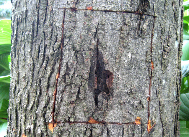
In late August 2006 I noticed several areas on the bark that were bleeding.
Curious as to what this bleeding was due to, I decided to carefully remove the bark at this location,
as shown below.
1
Using a hammer and chisel, I marked a rectangular area around the bleeding area.
Note that the surface bark seemed to have been eaten away at the center of the bleed area.
2
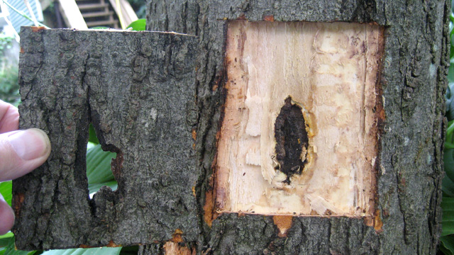
This picture shows the entire patch of bark removed from the tree.
The hole in the bark caused by the infection is clearly shown.
Furthermore, the infection has burrowed into the heartwood (xylem).
3
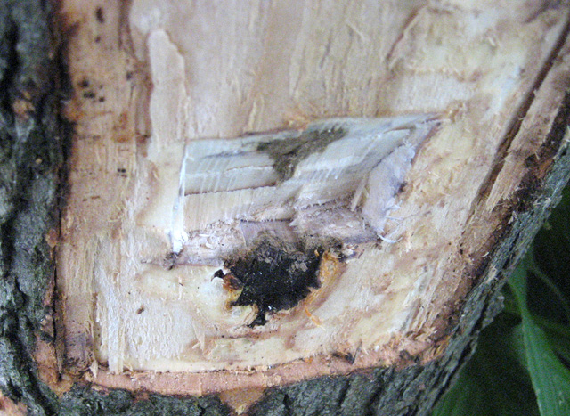
Curious as to what the cross-section of this canker looked like, I used a chisel
to dig through the canker to the center of the tree, as shown here.
As you can see, the canker shape is like an arrowhead pointing to the center of the tree.
The cause of this black, bleeding cankerous area is unknown. White Canker itself is not colored black.
Consequently, I suspect that White Canker weakened the tree to the point where a secondary fungal infection
took hold and burrowed into the tree at this point.
The tree continued its steady decline. About two years later (May 2008) the tree was about 80% dead.
But by then I had a better understanding of the symptoms of White Canker, so I again examined this tree
with the results shown below.
4
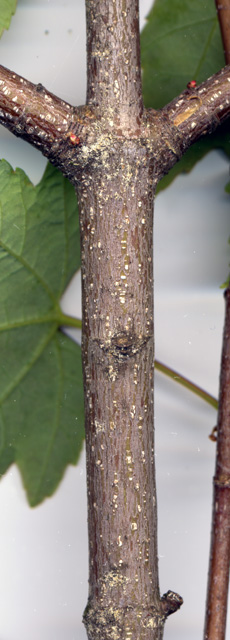
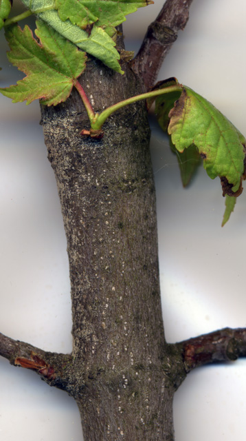
This picture was taken about 20 inches from the tip of the branch, and shows the spores
clustering at the branch junction and at the leaf scars, with very few spores in-between.
5
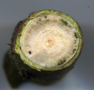
This area, about 3' from the branch tip, shows a large spore population.
At this location the spores are more diffuse along the branch.
But the total population of spores is larger, making this maple an efficient transmitter of white canker.
Notice also the new leaves sprouting near the heavy spore infestation.
The leaves are already misshapen and dying at the edges.
One is turning red, as if it were fall! (This picture was taken in mid-May, 2008)
6
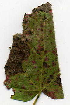
Here is a cross-section of this branch, showing the disease infection channels.
The following set of pictures were taken in late September 2008, after the spores had dispersed.
All the microscope pictures were taken at a magnification of 400x.
7
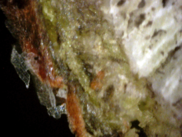
It's obvious when looking at this leaf that it is in very bad shape.It's parent
tree is already about 95% dead. The straight edge was made with a razor blade
for the purpose of making the cross-section views shown below.
8
Here there is white canker on the outer bark, although in this case the white
canker is actually transparent and looks like ice.
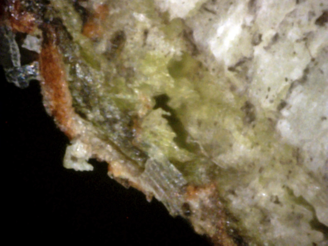
9
Here there is white canker under the bark and on the bark. Some is milk white
and some is transparent. The tubular form is almost always transparent.
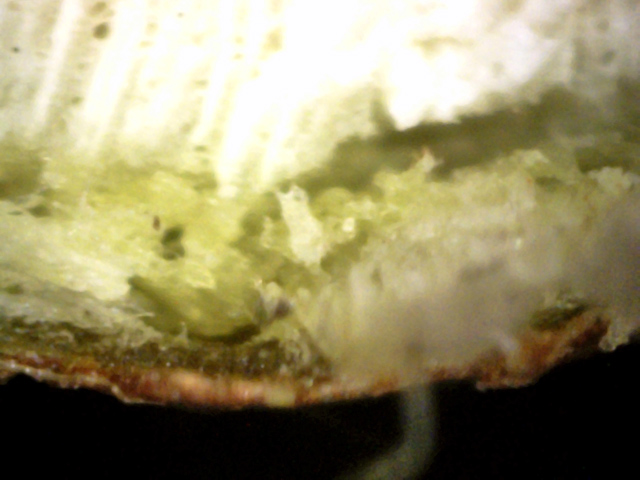
10
The inner bark is almost completely destroyed by canker. The gray blur in the
lower right is out of focus since it's too close to the camera.
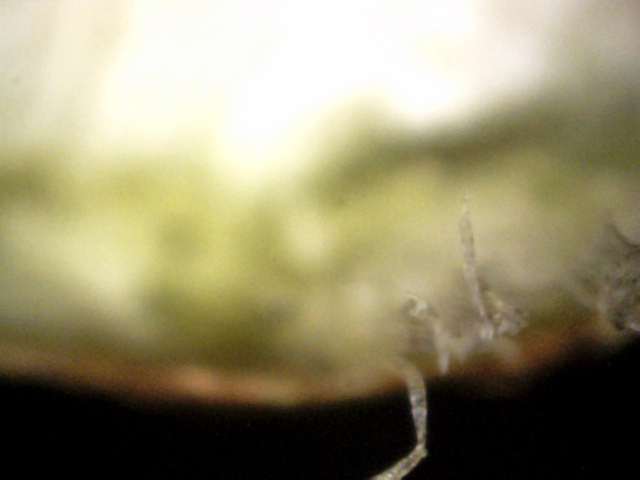
11
The gray blur in the prior picture is in focus here, and is shown to be composed
of transparent white canker hyphae.
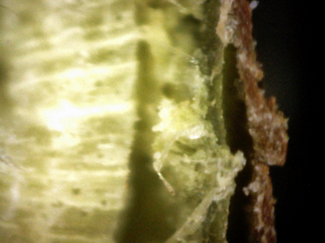
12
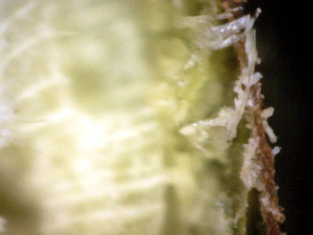
Here the white canker has eaten away all the inner bark, leaving a void. Note
where it is working away at the top of the picture. Also, the outer bark is
riddled with canker.
13
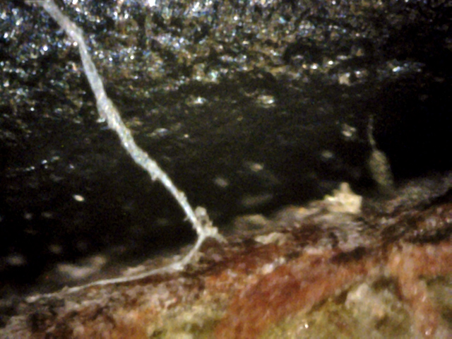
White canker hyphae are under and on the bark, sprouting white canker growths
along the hyphae.
 ↓
↓
↓
↓
↓
↓
14
Here is a long white canker hypha growing on the bark. It at first appears like
an icicle in the shape of a ribbon. If you look carefully, where the big bend
takes place, there is a small spore branching off (yellow arrow), and another
long growth (blue arrow) snakes over the bark to the canker just to the right
(red arrow).

15
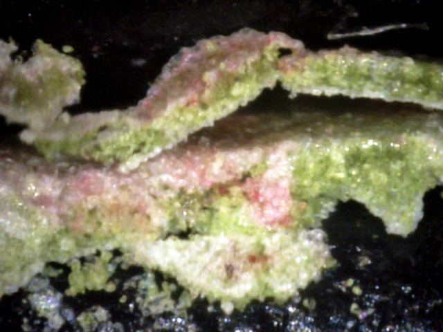
White canker under the leaf and in the leaf.
16
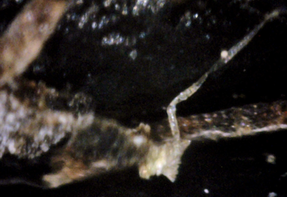
The canker infection has made a real mess of this leaf. The red color is
emerging fall maple leaf color.
 ↓
↓
17
A dead portion of th leaf that has a large blob of white canker material on the
bottom. This canker is spawning a hypha that starts off white and then turns
transparent. This hypha is budding off a young spore (red arrow).
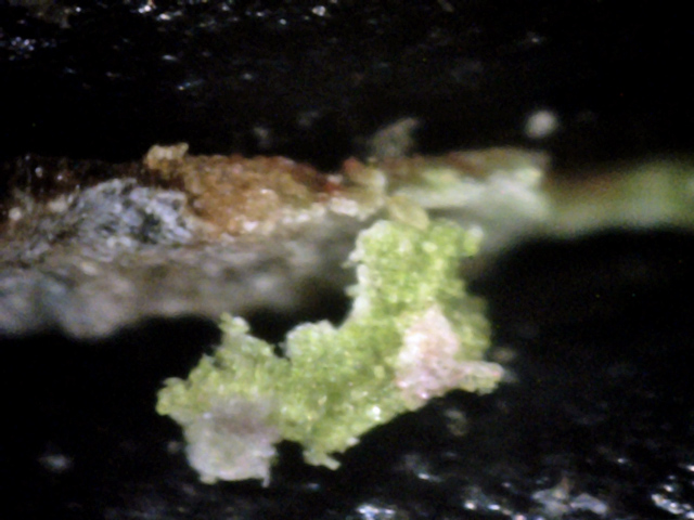
18
This piece of tissue torn from the leaf contains both white and gray canker
material.
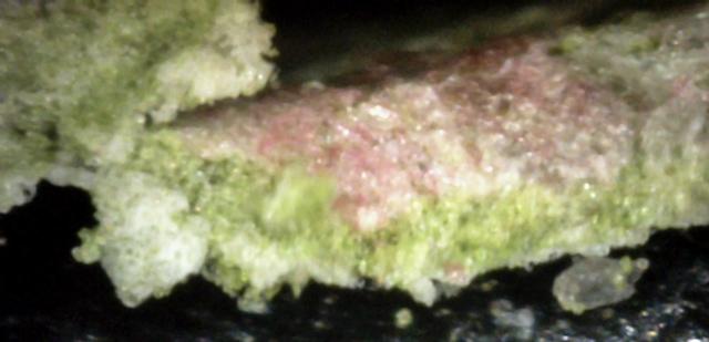
19
There is a white canker blob on the left, which looks kind of foamy. The pink
surface canker may be due to the leaf changing color in its stressed areas
first.
In Summary, this tree has a severe White Canker infection, to the point where
the tree is barely alive. All the typical signs of White Canker are present: distorted leaves,
bark splits, bleeding cankers, spores present on the bark, white canker material
within the leaves, and, most important, White Canker material just under the bark, choking
off the supply of nutrients to the tree.
These pictures are of a 30' red maple tree in my back yard, and are cuttings less than an hour old.
I happened to be doing some pruning on this tree and my eye caught these tan discolorations on the
branches that were about one-fourth to one-half inch in diameter.
All these branch discolorations covered only about 20 degrees of the trunk circumference, and were
at the bottom (ground facing) part of the branch.
Since this was late May, insect and other disease damage was almost non-existent.
1
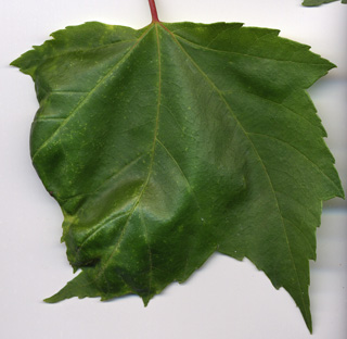
One branch had a twig sprouting from it, and that twig had about 5 leaves.
One of these leaves (shown here) had the typical white canker characteristics I'd seen
in prior years - puckering.
The puckered half of the leaf also had yellow spots.
2
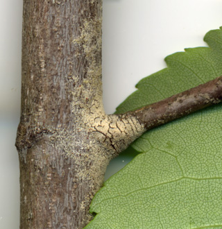
This picture, taken with a 2400 dpi scanner at the end of May, shows the junction of this leaf's twig to its branch.
The yellow substance is actually a massive quantity of tiny light yellow spores.
Even though the twig bark under the spores is very young, it is cracking.

3
As I continued to prune, I noticed many more of these yellow spore patches.
As in this picture, these dense patches were consistently at branch junctions and on the side
of the branch facing down. (While my unaided eye saw them as simply fuzzy patches, my 7 year old
grandson said he could discern individual spores.)
Since I couldn't believe these were all spores, I brought in numerous samples and scanned them
with a 400 power microscope. Sure enough - they were all spores, as the following pictures show.
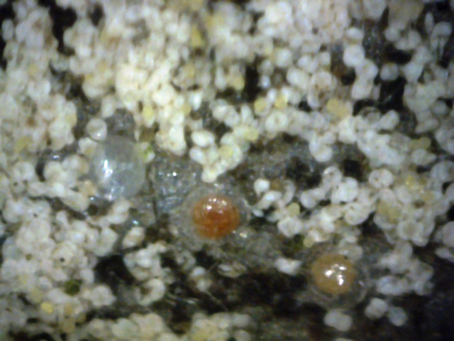
4
These are pictures of the yellow spore patches taken with a microscope.
At first glance, they may resemble a lot of teeth.
Or, the cleft separating their two lobes may remind one of 3D "Pac-men".
If you zoom in for a closer view, you'll see that each spore is milk-white in color, and
often has a small yellow object between the two lobes. This yellow object is like an egg yolk, and
seems to provide the food for the spore's growth. It shrinks in size as the spore matures.
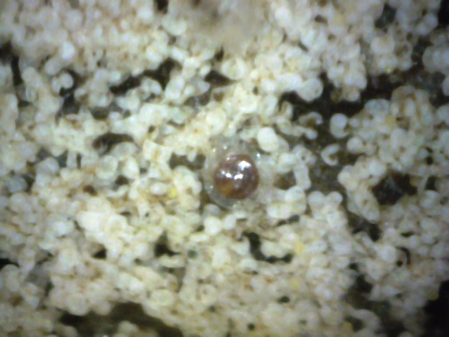
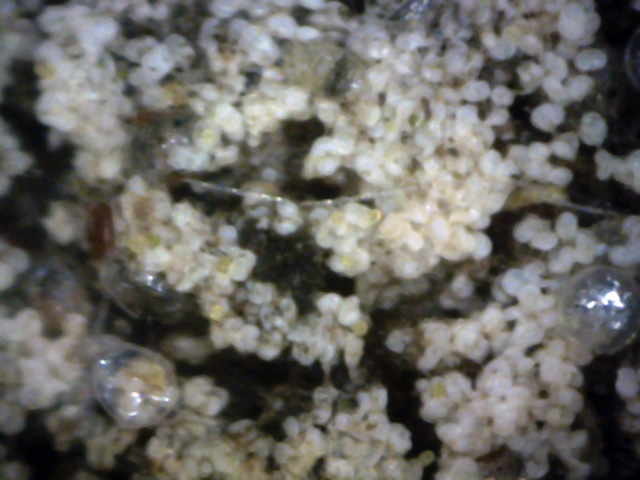
The several large gelatinous spherical objects among the spores may be
germinating spores from another host, since each seems to have a root-like
appendage. They may be the "pearls" in the following pictures.
5
6
The pictures shown above are of the spores on the bark of trees.
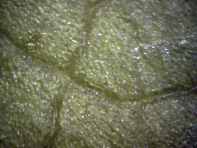
Here we use a 400x microscope to examine the upper and lower surface of the leaf shown in picture 1.
7
This is the top surface of the leaf.
The entire leaf looks pretty much like this - nothing much of interest for diagnostic purposes.
In fact, except for some yellow spots, you might think this was a healthy leaf.
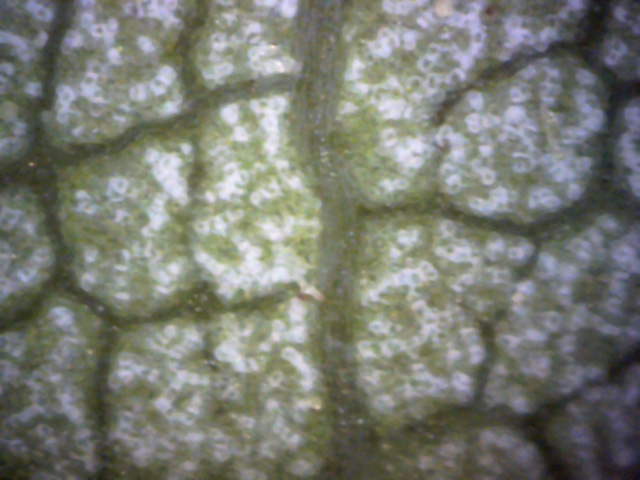
8
This is healthy tissue on the bottom of the leaf.
The white objects are stomata, through which the leaf breathes.
Once again, there is nothing visible that gives a clue that this leaf or branch is diseased.
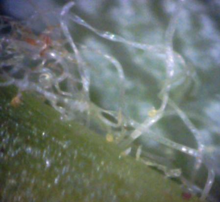
9
This picture shows the bottom of the leaf, near the stem.
There are several "yellow blobs" and numerous semi-transparent ribbon-like
filaments present.
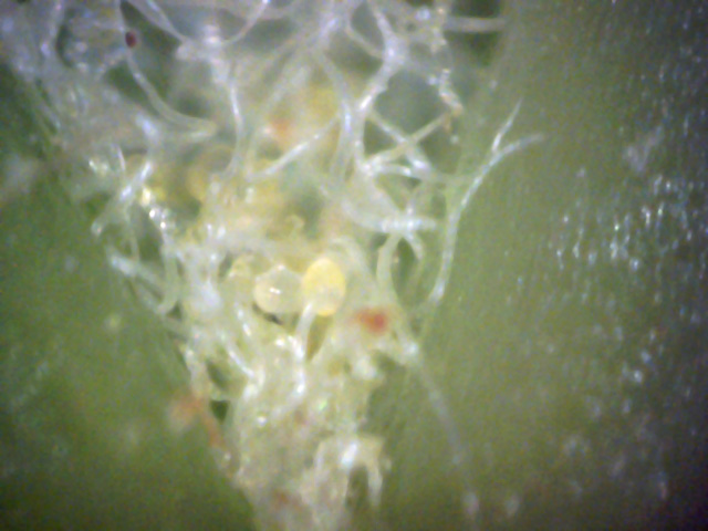
10
Since the spore density at branch junctions is high, I wondered if the same would be true at
leaf vein junctions on the bottom of the leaf.
As this picture shows, that indeed is true. There are two types of hyphae present - normal
leaf hyphae (round cross-section) and white canker hyphae (ribbon-like cross-section).
The yellow objects are embryonic white canker spores.
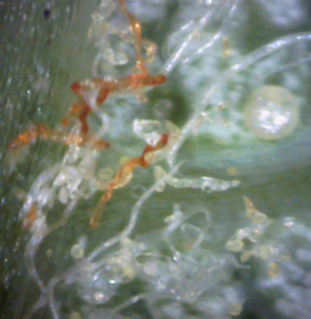
11
This picture was also taken near a leaf vein.
Some of the hypha are brown - possibly they are dying.
In addition to the small embryonic light yellow spores, there is a much larger tan globular
object present, which could be a germinating spore.
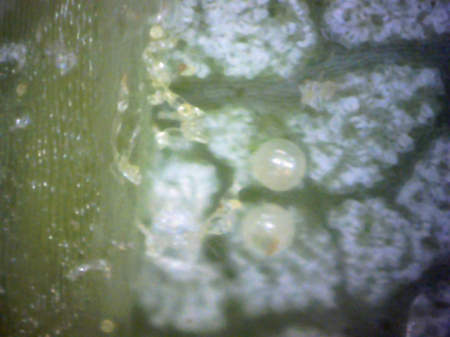
12
This picture too shows a leaf vein on the bottom of the leaf.
Hyphae are emerging from the vein and growing embryonic canker spores on them.
The two tan globular object may be germinating spores.
13
←
→
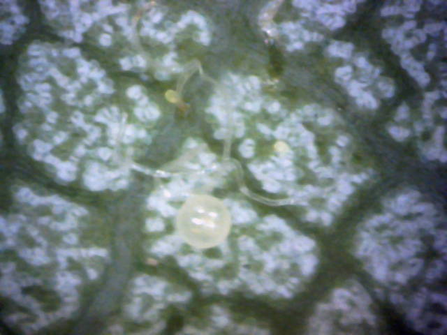
I included this leaf bottom picture because it shows that these yellow embryonic spores seem to have
a tiny attachment "tail" anchoring them to the tree tissue, and sometimes also have a
tiny black "eye" at the tip (see the black arrows).
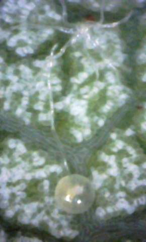
14
Another leaf bottom picture. Here again is one of those tan globular objects that looks like a pearl.
Look closely, and you can see the base of the pearl has a hypha which dives into the leaf right
next to an octopus-like hypha object. A embryonic yellow spore is also shown, and is connected to a
hypha.
15
Another "pearl" object. This picture clearly shows that this object is very likely
not an insect egg, since it has a hypha growing from its base, running along the leaf surface,
and then diving in right next to one of these "octopus-like" objects.
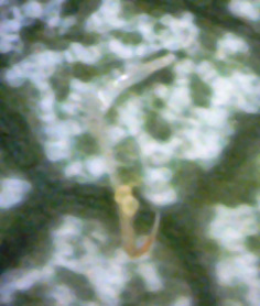
16
There are two young embryonic spores here - one anchored to the leaf and the other anchored
to a hypha.
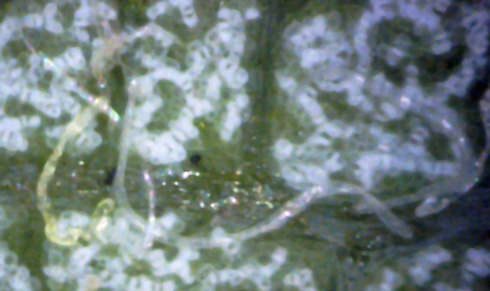
17
↑
↑
←
↓
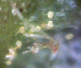
White canker hyphae tend to be transparent and ribbon-like in cross-section.
The two here, growing from black areas of the leaf vein (red arrows), have that characteristic.
The width to length ratio of these hyphae can be seen where a hypha twists (blue arrows).
It's rare to see a yellow hypha, as in the lower left of the picture.
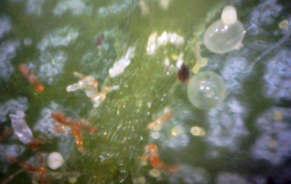
18
This part of the leaf vein seems to host a whole variety of white canker structures.
The "pearls" are there, embryonic spores are present, mature spores are present
live hyphae are present, and brown (probably dead) hyphae are also there.
In fact, you could say almost all the action takes place on or very near the leaf veins!
After reviewing these pictures, some interesting conjectures can be made:
- Except for leaf puckering, a microscopic examination of the top side of a leaf gives almost no clue as to the presence of this disease.
- All the microscopic leaf evidence for this disease appears as light yellow spores on the leaf bottom, near the diseased (puckered) area.
- The bark may contain a huge number of both white and light yellow small spores (leaf bottoms contain no white spores).
- The yellow spores on the leaf seem to take root in it via a small "tail".
- "Pearls" often have a hypha coming from them which terminates near a "spider-like" object.
- Some filaments are brown in color rather than transparent. These may be filaments that have died.


 ↓
↓
↓
↓
↓
↓

 ↓
↓


